Psychology
The Neuropsychology of Religious and Spiritual Experience
Religious and spiritual experiences are rooted in the brain's evolutionary and structural architecture. Examining their neurophysiological correlates and cognitive operators offers a model for understanding these experiences biologically and their connection to psychological well-being.

Religious and spiritual experiences, such as meditation, prayer, and rituals, have been described in biomedical, psychological, anthropological, and religious writings. The history of some of these specific descriptions and religious texts goes back several thousand years. Recently, there has been a growing increase in the number of studies exploring the neurophysiological and physiological correlates of such experiences. From an evolutionary perspective, such experiences may have become possible with the development of various brain structures in early primates, and ultimately in Homo sapiens...
.
The Neuropsychology of Religious and Spiritual Experience
Andrew B. Newberg
Stephanie K. Newberg[1]
Translated by Mohsen Zandi (Researcher in the Psychology of Religion)
Religious and spiritual experiences, such as meditation, prayer, and rituals, have been described in biomedical[2], psychological, anthropological, and religious writings. The history of some of these specific descriptions and religious texts goes back several thousand years. Recently, there has been a growing increase in the number of studies exploring the neurophysiological[3] and physiological correlates of such experiences. From an evolutionary perspective, such experiences may have become possible with the development of various brain structures in early primates, and ultimately in Homo sapiens,[4]. It is thought that the emergence of "religiotropic"[5] brain mechanisms in Homo sapiens historically coincided with the sudden growth of religious traditions, which have continuously found their way into human societies since prehistoric times. In light of this evolutionary model, the neurobiological and neuropsychological[6] correlates of religious and spiritual experiences have gradually been identified. Also, considering other related research in neurobiology,[7] a more complex pattern of neuropsychological events during religious and spiritual experiences can be obtained. More precisely, brain function can be examined with regard to its interconnections with other parts of the body that are affected by the autonomic nervous system as well as the neuroendocrine system. Examining the relationship between cognitive processes in the brain and the autonomic nervous system may lead to a more comprehensive understanding of the many varieties of spiritual experiences, which range across a spectrum from "awe" to intense mystical states. Consequently, current works can be used as a foundation for developing a neuropsychological model to guide future research on the neurobiology of religious and spiritual experiences. The most advanced brain imaging methods, which can examine various neurotransmitter networks, as well as other physiological measures, can be employed in investigating brain function during experiences such as meditation, supplication and prayer, and ritual experiences.
This chapter analyzes the neuropsychology of religious and spiritual experiences and includes a brief examination of the phenomenological aspects of such experiences, as well as the synthesis of existing data to develop a comprehensive model that can provide a foundation for future analyses of the biological roots of these experiences, and the relationship between these experiences and psychological well-being.
The Evolution of the Brain and Spiritual Experience
The evolutionary process has led to the emergence of complex neural connections that exist in the cerebral hemispheres. Moreover, higher centers in the brain are connected with more primitive structures such as the limbic system. Overall, the brain evolved its complexity in the evolutionary process to provide humans with advanced abilities for representing the order of the external environment and solving the cognitive problems necessary for survival. In addition to purely cognitive aspects, the evolution of the brain led to human socialization. This ability to create family units, groups, communities, and societies conferred a tremendous evolutionary advantage. Therefore, our question here is: how do these evolutionary changes in the brain lead to the emergence and development of spiritual experiences, religion, rituals, and rites?
Functionally, the brain can be divided into several fundamental cognitive functions (d’Aquili, 1978, 1983, 1986). We have previously called these functions "cognitive operators"[8]. The term cognitive operator refers only to the neurophysiological mechanisms that underlie certain broad areas of cognitive function. Thus, these operators do not exist in the strict sense of the word; rather, (assuming them) is useful for examining the overall function of the brain. The concept of cognitive operators is similar to the more widely used concept of modules[9] or cognitive units. However, by assumption, cognitive operators refer to the brain's more general functions. These cognitive operators include the abstraction of the general [or whole] from particulars, the perception of causality in external reality, the perception of spatial or temporal contiguity in external reality, and the ordering of fragments of reality into a causal chain. This latter function is probably what creates explanatory models[10] of the external world; whether the explanation is scientific or mystical and esoteric. There is no room here to explain the details of the physiological substrates and neuroanatomical networks[11] of all these operators. Nevertheless, a brief explanation of a few operators can be useful in elucidating the neuropsychology of religious and spiritual experiences.
The causal operator is involved in the causal ordering of elements of reality that are abstracted from sensory perceptions (d’Aquili, 1978). The causal operator derives its function from the inferior parietal lobule in the left hemisphere, the anterior convexity of the frontal lobes, mostly in the left hemisphere, and from bilateral intracerebral connections (Luria, 1966; Pribram, 1973). The causal operator is thought to have fundamental importance in the generation of religious and spiritual experiences and concepts (d’Aquili, 1978). This operator organizes any given fragment of reality into a subjective causal continuum and traces it back to its initial station. Reflecting on the (presumably) pervasive human characteristic of assigning a cause for every piece of reality, it can be said that even if the initial station of the aforementioned causal chain is not obtained through sensory data, the causal operator automatically generates that initial causal station (d’Aquili & Newberg, 1993). Western science does not accept the postulation of an initial station or first cause for fragments of reality unless that first cause is either observed or can be directly inferred from [sensory] observation. In everyday (non-scientific) life, the causal operator simply constructs an initial station or first cause for every piece of reality. We suspect that when no scientific causal explanation or observation is available for a piece of reality, gods, [higher] powers, spirits, or some other causal structures are automatically generated by the causal operator[12] (d’Aquili & Newberg, 1997). If this is the case, when our observations do not reveal the initial station, the causal operator perhaps spontaneously intervenes and generates a primary causal terminus for external reality.
If it is true that the causal operator necessarily analyzes reality, then humans have no choice but to construct myths filled with personified forces to explain the world around them. Myths may well be social in nature, or, based on dreams, fantasies, or other imaginative aspects of the individual, a phenomenon with an individual coloring. Nevertheless, insofar as humans are aware of the possibility of their existence in the face of the unstable and unreliable world around them, they create myths to identify their path and place in the universe and to adapt to their surrounding habitat. Hence, they create gods, spirits, demons and angels, or other sources of personified power with whom they can make contracts and transact, in order to thereby gain control over their unstable and unreliable ecosystem.
The second operator that we believe holds particular importance in spiritual experience is the holistic operator[13]. The proposed holistic operator allows the human being to view reality in the form of a whole or gestalt[14] and enables the abstraction of a broader contextual framework[15] from the parts or individuals. The causal operator is, presumably, located in the parietal lobe of the right hemisphere (or the non-dominant hemisphere),[16] and more precisely, in the superior posterior parietal lobule and adjacent areas, which we now know are involved in producing a gestalt understanding of various sensory data and abstract concepts (Bogen, 1969; Gazzaniga & Hillyard, 1971; Nebes & Sperry, 1971; Sperry, Gazzaniga, & Bogen, 1969). It is also interesting to note that this area is situated opposite an area in the left hemisphere that provides the neuroanatomical substrate[17] for logical-grammatical functions. Consequently, the right parietal lobe mediates a holistic approach to things, and the left parietal lobe mediates more reductionist[18] processes. We will examine below how these various structures, and related structures, might be involved more specifically in religious and spiritual experiences.
Methods of Attaining Spiritual Experiences
In examining the neuropsychological model of religious and spiritual experiences, it is crucial to dissect how such experiences are attained. We suggest there are two general categories in the methods of achieving such experiences: group rituals,[19] and individual contemplation,[20] such as prayer or meditation. Phenomenological analysis of these two types of practice shows that they share a common genus (though not in intensity and severity): 1) periodic emotional discharges that involve a subjective sense of awe, peace, tranquility, or ecstasy (self-transcendence); and 2) varying degrees of unitary experience, which correspond to the aforementioned emotional discharges (d’Aquili & Newberg, 1993). These unitary experiences consist of a decreased sense or awareness of the boundary between the “self” and the external world (d’Aquili, 1986; d’Aquili & Newberg, 1993; Smart, 1958, 1967, 1969; Stace, 1961). The latter dimension can also lead to a sense of unity among other individuals within the perceptual field, thereby generating a sense of communitas[21]. Ultimately, unitary experiences can result in the dissolution of all boundaries of discrete and discontinuous existence, consequently bringing about a state of undifferentiated oneness (or what we have called Absolute Unitary Being[22]) (AUB; d’Aquili & Newberg, 1999). It should be noted that group ritual experiences and individual meditation overlap to some extent; insofar as each can play a role in the genesis of the other. Indeed, it may be that ritual ceremonies create the “average” person[23] who attains mystical and religious experience (“average,” in distinction from those who regularly and frequently engage in intense contemplation, such as monks and highly devout ascetics). This is by no means to say that the mystical and contemplative experience is indifferent to the effects of ritual ceremonies. It is precisely because of the intense unitary experiences resulting from meditation that the susceptibility of mystics to ritual ceremonies may, in practice, be greater than that of the average person; although this has yet to be proven. One might conclude that ritual ceremonies, at their most effective, are an immensely powerful technique (whether for good or ill). Also, due to its inherently public dimensions, it usually has far greater social significance than individual meditation or contemplation. Although meditation and contemplation can generate more intense and extensive unitary states (which have been likened to the relatively brief flashes produced by group ritual), they are almost always solitary and reclusive experiences.
It seems that ritual practices are morally neutral techniques; that is, they can be used for both constructive, positive ends and negative, destructive ones. Thus, ritual, relying on a myth that both contains and expresses the ritual practices, can expand or diminish the structural aspects of a community[24], and can provoke or reduce aggressive behavior. Employing Turner's (1969) theory of communitas (as a powerful experience of social unity that occurs most often as a result of ritual practices), we can say that if a myth finds its embodiment in a ritual that determines the experience of unity for a specific group or tribe, then the result is merely unity and tribal communitas[25]. It is a true statement that by unifying the experience created by ritual, aggression and transgression within the group are minimized or reduced to zero. However, even if intra-group transgression and aggression are reduced, this experience of intra-group unity may "only" serve to emphasize the group's integration in distinction and opposition to other groups, and consequently, lead to an increase in inter-group aggression. Of course, sometimes the scope of the myth and its ritual embodiment expands to include all members belonging to a religion, a nation-state, an ideology, all human beings, or all of reality. Clearly, the greater the scope of what is intended in the unitive experience, the more aggressive behavior diminishes. In fact, if it is a true statement that ritual practices can sometimes flesh out the myth of the unity of all beings, then the individual performing such a ritual may experience a fleeting sense of the unity of all persons. Such a mythic-ritual experience[26] is something akin to meditative states, such as Bucke's (1961) cosmic consciousness or even AUB[27]. However, unfortunately, in ethnographic experiences, such a prominent scope of unity in group rituals has been very rare.
A Neuropsychological Model of Religious and Spiritual Experiences
The model described below is a more detailed model than the one previously described, which now incorporates recent studies from brain imaging, neurochemistry,[28] hormonal, and physiological research (Newberg & Iversen, 2003). The goal of this model is to build a foundation through which the many varieties of religious experiences and practices can be examined and compared. As Figure 11.1 shows, this model begins with the prefrontal cortex, and describes a number of complex interactions with the thalamus, the superior posterior parietal lobe,[29] the limbic system, and the autonomic nervous system. Also, a number of excitatory and inhibitory neurotransmitters[30] can be shown to play a role in such practices and experiences. Dopamine, serotonin, acetylcholine, and several other molecules can be linked to various phenomenological aspects of such experiences; all of these are analyzed in this model. The specific mechanisms may differ somewhat based on the practice, ritual, tradition, or specific individual. However, by focusing on the phenomenology of such experiences, this model provides information about the variety of experiences, whether sensory, cognitive, or emotional, that can be associated with religious and spiritual experiences. Since a more or less extensive amount of data was available, this model was primarily created by employing data obtained from research that focuses mostly on meditative practices. This model may possibly be applied to a very wide variety of practices and experiences.
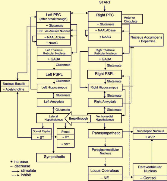
Prefrontal and Cingulate Cortex Activation[31]
Most meditative practices, prayer, or other contemplative acts require a degree of sustained, uninterrupted attention. This sustained maintenance of attention can be achieved by focusing on the image of an object, a mantra [hymns in Hinduism], a prayer, or some other spiritual focus. Brain imaging research indicates that voluntary, deliberate activities and tasks requiring the uninterrupted maintenance of attention are initiated by activity in the prefrontal cortex[32] (PFC), particularly in the right hemisphere (Frith, Friston, Liddle, & Frackowiak, 1991; Ingvar, 1994; Pardo, Fox, & Raichle, 1991; Posner & Petersen, 1990). The cingulate gyrus[33] has also been shown to cooperate with the PFC in attentional focus, likely (Vogt, Finch, & Olson, 1992). Consequently, since spiritual practices require intense concentration of attention, it seems a valid hypothesis that the model intended for meditation begins with the activation of the prefrontal cortex (particularly in the right hemisphere), as well as the cingulate gyrus. This idea has been supported by the increased activity observed in these regions in several brain imaging studies on various types of voluntary meditation; including studies conducted in our own laboratory in which eight Tibetan Buddhist meditators were examined at baseline and during meditation (Newberg et al., 2001). Quantitative analysis showed increased activity in the prefrontal cortex bilaterally (though more so in the right hemisphere) and the cingulate gyrus during meditation. Thus, it appears that meditation begins with the activation of the prefrontal and cingulate cortex, which are associated with the volitional clarification of mental thoughts or focusing on an object. However, a positron emission tomography[34] (PET) study on a guided form of meditation did not show increased prefrontal activity; although, a recent study comparing internally generated versus voluntary speech showed decreased anterior activity during externally guided speech production (Crosson et al., 2001). Consequently, prefrontal and cingulate activation may be associated with the volitional aspects of meditation.
Thalamic Activation as Part of the Attentional Network
The thalamus is a major relay station in the brain that links other structures, and also transmits higher-order processing to parts of the brain involved in emotion; and finally, the thalamus regulates various physiological processes. Several animal studies have shown that the prefrontal cortex innervates the reticular nucleus of the thalamus during arousal[35] ( Phillipson, 1988&Cornwall); particularly as part of a more integrated attentional network (Portas et al., 1998). Such arousal may be accomplished through the production and distribution of the excitatory neurotransmitter glutamate by the prefrontal cortex, which prefrontal cortex neurons use for transmission and communication among themselves, and for innervating other brain structures (Cheramy, Romo & Glowinski, 1987 ). The thalamus itself governs the flow of sensory data toward cortical processing areas through its interactions with the lateral and lateral posterior geniculate nuclei[36] and is thought to also use the glutamate network to activate neurons in other structures (Armony & LeDoux, 2000). We know that the lateral geniculate nucleus receives raw visual data from the visual apparatus and sends it via a specific pathway to the striate cortex[37] for processing. The lateral posterior nucleus of the thalamus provides sensory data to the posterior superior parietal lobule[38] (PSPL), which needs them for spatial orientation (Bucci, Conley, & Gallagher, 1999).
During arousal, the reticular nucleus secretes the inhibitory neurotransmitter gamma-aminobutyric acid (GABA) toward the lateral posterior and geniculate nuclei, which cuts off input to the PSPL and visual foci in proportion to the arousal of the reticular nucleus (Destexhe Contreras, & Steriade, 1998). During meditation, due to increased activity in the PFC, particularly in the right hemisphere, there should theoretically be a concomitant increase in the activity of the thalamic reticular nucleus. While brain imaging studies of meditation have not had a solution for distinguishing the reticular nuclei, our single recent single-photon emission computed tomography[39] (SPECT) study showed an overall increase in thalamic activity that was proportional to activity levels in the PFC. This is consistent with (but does not confirm) a specific interaction between the PFC and the reticular nuclei. If right-hemisphere PFC arousal causes increased activity in the reticular nucleus during meditation, then the result may be a reduction in the sensory input entering the PSPL. Several studies have shown an increase in serum GABA [GABA] during meditation, which is thought to reflect increased central GABA activity (Elias, Guich, & Wilson, 2000). This functional deafferentation[40] associated with increased GABA means that fewer distracting and disturbing external stimuli reach the visual cortex and PSPL; a phenomenon that occurs with the increased sense of focus during meditation.
It should also be recalled that the dopaminergic[41] network, via the basal ganglia[42], participates in regulating the glutamatergic[43] network and the interaction between the prefrontal cortex and subcortical structures. A recent PET [positron emission tomography] study measured dopamine tone during Nidra Yoga[44] meditation and showed a significant increase in dopamine levels during the act of meditation (Kjaer et al., 2002). Kjaer and colleagues hypothesized that this increase may be associated with a suppression of cortical and subcortical interactions, leading to an overall reduction in readiness for action; which is also associated with this particular type of meditation. Future studies need to carefully examine and clarify the role of dopamine during meditative practices, as well as the interactions between other neurotransmitter networks.
Deafferentation of the PSPL
Research has shown that the PSPL is involved in the analysis and integration of high-order visual, auditory, and somatic aesthetic data (Adair, Gilmore, Fennell, Gold & Heilman, 1995; see also Joseph, 1990). As we mentioned earlier, this structure is thought to play a fundamental role in both holistic and reductionist cognitive processes. The PSPL also participates in a complex attentional network involving the PFC and the thalamus (Fernandez-Duque & Posner, 2001). By receiving auditory and visual input from the thalamus, the PSPL is empowered to help create a three-dimensional image of the body in space, generate a sense of the spatial coordinates in which the body is oriented, distinguish between objects, and influence the perception of objects that can be directly grasped, used, and manipulated (Mountcastle, Motter, & Anderson, 1980; Lynch, 1980). These functions of the PSPL are perhaps crucial for distinguishing between the self and the external world. We should also note that recent research suggests the superior temporal lobe may play a more fundamental role in the spatial representation of the body; although this finding has not been confirmed by other reports (Karnath, Ferber, & Himmelbach, 2001). In any case, the question remains: what is the true relationship between the parietal and temporal lobes in terms of spatial representation?
Nevertheless, we believe that the deafferentation of these orientation areas of the brain holds an important concept in the physiology of meditation. For example, if deafferentation of the PSPL occurs through the effects of the gamma-aminobutyric acid network[45] of the reticular nucleus, the person may begin to lose their usual ability for spatial self-recognition. This idea is supported by clinical findings in patients with Balint's syndrome[46], where parietal lobe damage causes significant difficulty for sufferers in recognizing themselves in three-dimensional space. Of course, the effects of meditation may be more selective and not destroy the "sense of self"; rather, they may alter its perception[47]. Furthermore, deafferentation of the PSPL is corroborated by two imaging-based studies showing reduced activity in this area during intense meditation (Newberg et al., 2001; Herzog et al., 1990–1991). Our SPECT study also showed a correlation between increased activity in the thalamus and decreased activity in the PSPL. The implication of this is that as the individual's PFC activity increases, the deafferentation of the PSPL intensifies. Thus, a researcher might hypothesize that the more intense and deeper the concentration, the greater the likelihood that the person will eventually attain unitary states.
Excitation of the Hippocampus and Amygdala During Spiritual Practices
In addition to complex cortical-thalamic activity, one can expect that meditative and spiritual practices also alter limbic system activity; especially since the stimulation of limbic structures is associated with experiences similar to those described during these practices (Fish, Gloor, Quesney, & Olivier, 1993; Saver & Rabin, 1997). The hippocampus acts to regulate and modulate cortical arousal and responsiveness through its extensive and rich interconnections with the prefrontal cortex, neocortical regions,[48] the amygdala, and the hypothalamus (Joseph, 1990). It has been found that hippocampal stimulation reduces cortical arousal and responsiveness; however, if cortical arousal is initially at a low level, then hippocampal stimulation usually increases cortical activity (Redding, 1967). The ability of the hippocampus to excite or inhibit neuronal activity in other structures may be based on glutamate and GABA networks, respectively (Armony & LeDoux, 2000). Our idea in our neuropsychological model of meditation is that during meditation there is a partial deafferentation of the right hemisphere's PSPL. This deafferentation, also due to the inverse regulation of the hippocampus relative to cortical activity, can lead to right hippocampal stimulation. Furthermore, if there is direct and simultaneous stimulation of the right hippocampus via the thalamus (as part of the known attentional network), and it is influenced by glutamate, we believe a powerful potentiation of right hippocampal stimulation then occurs. Right hippocampal activity may ultimately enhance the PFC's stimulatory function on the thalamus via the nucleus accumbens,[49] which is poised to interrupt neural input from the PFC to the thalamus through the neuromodulatory effects of dopamine (Newman & Grace 1999).
The hippocampus exerts a significant influence on the amygdala, such that they complement each other and interact in generating attention, emotion, and specific types of mental imagery (Joseph, 1990). It appears that much of the prefrontal regulation of emotion is carried out through the hippocampus and its connections with the amygdala (Poletti & Sujatanond, 1980). Because of this bidirectional interaction between the amygdala and the hippocampus, we suspect that right hippocampal stimulation during meditation likely stimulates the lateral amygdala as well. Functional magnetic resonance imaging (fMRI)[50] results by Lazar and colleagues (2000) support the belief in increased activity in parts of the amygdala and hippocampus during meditation.
Hypothalamic and Autonomic Nervous System Changes
We know that the hypothalamus is extensively interconnected with the limbic system. Research has shown that stimulation of the right lateral amygdala leads to stimulation of the ventromedial part [nucleus] of the hypothalamus, with subsequent stimulation of the peripheral parasympathetic system (Davis, 1992). Increased parasympathetic activity is associated with the initial subjective feeling of relaxation, and ultimately, with a deeper sense of stillness and tranquility. Furthermore, parasympathetic system activation is the cause of decreased heart rate and respiratory rate. All of these physiological responses have been observed during meditation (Jevning, Wallace, & Beidebach, 1992).
For example, when the respiratory and heart rates decrease, the nucleus paragigantocellularis of the medulla[51] ceases to function in order to innervate the locus coeruleus[52] (LC) of the pons. The locus coeruleus produces and distributes norepinephrine (NE) (Foote, 1987); a neuromodulator that increases the receptivity of brain regions to sensory inputs by amplifying strong stimuli, while simultaneously filtering out weaker excitations and cellular "noise"[53] that falls below the threshold of excitation (Waterhouse, Moises, & Woodward, 1998). Reduced stimulation of the locus coeruleus leads to decreased levels of norepinephrine (Van Bockstaele & Aston-Jones, 1995). It has generally been observed that the disruption of catecholamine products such as norepinephrine and epinephrine in urine and plasma decreases during meditation (Walton, Pugh Gelderloos, & Macrae, 1995), which may simply indicate a systematic shift in the balance of the autonomic nervous system. However, this is not inconsistent with a cerebral[54] reduction in norepinephrine levels. Our proposed model suggests that during meditation, decreased firing of the nucleus paragigantocellularis reduces its innervation of the locus coeruleus; which we know in turn supplies norepinephrine to the PSPL and the lateral posterior nucleus (Foote, 1987). Hence, the reduction in norepinephrine diminishes the impact of sensory input on the PSPL by contributing to its deafferentation.
Furthermore, the locus coeruleus delivers less norepinephrine to the paraventricular nucleus[55] of the hypothalamus. The paraventricular nucleus of the hypothalamus constantly secretes corticotropin-releasing hormone (CRH) in response to neural stimulation by norepinephrine from the locus coeruleus (Ziegler, Cass, & Herman, 1999). This CRH stimulates the anterior pituitary to release adrenocorticotropic hormone[56] (ACTH) (Livesey, Evans, Mulligan, & Donald, 2000). ACTH in turn stimulates the adrenal cortex to produce cortisol, which is one of the body's stress hormones (Davies, Keyon, & Fraser, 1985). The reduction of norepinephrine from the locus coeruleus during meditation likely decreases CRH production by the paraventricular nucleus, and ultimately cortisol levels. Most research has found that urinary and plasma cortisol levels decrease during meditation (Jevning, Wilson, & Davidson, 1978; Sudsuang, Chentanez, & Veluvan, 1991), a finding that supports the theory that there is an overall decrease in cortisol secretion. This also has implications for the relationship between meditative practices and stress reduction, as cortisol is consistently regarded as the primary stress hormone.
The drop in blood pressure associated with parasympathetic activity is expected to calm the arterial baroreceptors during meditative practices, which in turn leads the caudal ventrolateral medulla[57] to reduce gamma-aminobutyric acid (GABA) inhibition[58] of the supraoptic nucleus of the hypothalamus. Under certain conditions, this lack of inhibition can stimulate the supraoptic nucleus to release the vasoconstrictor hormone arginine vasopressin[59] (AVP), causing arteries to constrict and blood pressure to return to normal levels (Renaud, 1996). AVP has also been shown to contribute to the general maintenance of positive affect (Pietrowsky, Braun, Fehm, Pauschinger, & Born, 1991), reduce perceptions of arousal and fatigue, and significantly improve the consolidation and enhancement of new memories and learning (Weingartner et al., 1981). Indeed, plasma AVP has been shown to increase markedly during meditation (O’Halloran et al., 1985). Thus, an increase in AVP could lead to a reduction in the subjective feeling of fatigue and an increase in the sense of arousal. It could also help enhance the meditator's memory of the experience they are undergoing, and perhaps provide an explanation for the subjective phenomenon of why spiritual and meditative experiences are remembered, recalled, and described in passionate terms.
PFC Influences on Other Neurochemical Networks
As the spiritual practice continues, the PFC activity associated with the sustained, diligent will to focus attention must also continue. Generally, when PFC activity increases, it produces rising levels of synaptic glutamate released in the brain. Increased glutamate can stimulate the arcuate nucleus of the hypothalamus to release beta-endorphin[60] (BE) (Kiss, Kocsis, Csaki, Gorcs, & Halasz, 1997). BE is an opioid primarily produced by the arcuate nucleus of the medial hypothalamus[61] and distributed to subcortical regions of the brain (Yadid, Zangen, Herzberg, Nakash, & Sagen, 2000). We know that BE reduces respiration, diminishes fear and pain, and produces feelings of joy and euphoria (Janal, Colt, Clark, & Glusman, 1984). Those effects described from the meditative state may indicate the release of some BE associated with increased PFC activity. Research has found that meditation disrupts the daily cycles of BE and ACTH, although it does not affect the daily cycles of cortisol (Infante et al., 1998). However, it is thought that BE is not the sole mediator of such experiences during meditation, because the mere reception of morphine-related substances does not produce equivalent mystical experiences. Also, a very limited study showed that blocking opioid receptors with naloxone[62] had no effect on this experience, or on the electroencephalogram (EEG) associated with meditation (Sim & Tsoi, 1992).
Glutamate activates N-methyl-D-aspartate receptors[63] (NMDAr); however, excess glutamate can destroy these neurons through the process of excitotoxicity[64] (Albin & Greenamyre, 1992). We suspect that if glutamate levels approach excitotoxic concentrations during intense meditative states, the brain may limit the production of the acidic dipeptidase N-acetylated-alpha-linked[65] (which converts the endogenous anti-NMDAr N-acetyl aspartyl glutamate[66] to glutamate) (Thomas, Vornov, Olkowski, Merion, & Slusher, 2000). Increased NAAG protects cells from excitotoxic damage. However, there is an important side effect; the NMDAr-inhibiting NAAG is functionally comparable to the dissociative hallucinogens ketamine,[67] phencyclidine, and nitrous oxide[68] (Jevtovic- Todorovic, Wozniak, Benshoff, & Olney, 2001). These NMDAr antagonists produce various states whose characteristics can be described in terms of schizophrenic experiences or mystical experiences such as out-of-body experiences,[69] and near-death experiences (Vollenweider et al., 1997).
Autonomic Nervous System Activity[70]
In the early 1970s, Gellhorn[71] and Kiely[72] presented a model of the physiological processes involved in meditation that was based almost solely on autonomic nervous system (ANS) activity; although somewhat limited, it indicated the importance of the ANS during such experiences (Gellhorn & Kiely, 1972). These authors' idea was that intense stimulation of either the sympathetic or parasympathetic system, if sustained, could eventually lead to the simultaneous discharge of both systems (something that could be considered an "advance" of the other system). Several studies have shown a predominance of parasympathetic activity during meditation, which is associated with decreased heart rate and blood pressure, respiratory rate, and reduced oxygen metabolism (Travis, 2001). Nevertheless, a recent study of two independent meditation techniques suggested the co-activation of both the sympathetic and parasympathetic systems by demonstrating increased heart rate variability during meditation (Peng et al., 1999). The hypothesis of this research was that increased heart rate variability reflects the activation of both arms of the autonomic nervous system. This view also aligns with the specific description of meditative states as phenomena involving feelings of profound calm and stillness, as well as remarkable alertness. Furthermore, the theory of reciprocal activation of both arms of the autonomic nervous system is consistent with recent advances in the study of autonomic interactions (Hugdahl, 1996).
Serotonergic System Activity[73]
Autonomic nervous system arousal can lead to intense stimulation of structures in the lateral hypothalamus and the medial forebrain bundle[74], which we know produce feelings of ecstasy, euphoria, and bliss whenever directly stimulated (Olds & Forbes, 1981). Stimulation of the lateral hypothalamus can also lead to changes in serotonergic network activity. In fact, several studies have shown that after meditation, the breakdown of serotonin (ST) metabolites in urine increases markedly, indicating an overall increase in ST during meditation (Walton et al., 1995). Serotonin is a neuromodulator that densely supplies the visual centers of the temporal lobe; there, serotonin potently influences the flow of visual associations generated by this area (Joseph, 1990). The dorsal raphe cells produce and distribute ST when stimulated by the lateral hypothalamus (Rasmussen,1987&Aghajanian, Sprouse), as well as when activated by the prefrontal cortex (Juckel, Mendlin & Jacobs, 1999). A relative increase in ST levels correlates with positive affect, whereas low ST levels often indicate depression (Van Praag & De Haan, 1980). This link has been clearly demonstrated regarding the effects of selective serotonin reuptake inhibitor drugs,[75] which are widely used to treat depression. We should also recall that several clinical studies have found that meditative and spiritually-oriented practices can reduce rates of depression or relapse into depression. The link between spiritual practices and reduced depression confirms the role of serotonin in spiritual practices.
Nevertheless, when cortical ST receptors (especially in the temporal lobes) are activated, the stimulation can lead to hallucinogenesis. Tryptamine psychedelics, such as psilocybin and LSD, appear to make the most use of this mechanism to produce their astonishing visual associations (Aghajanian & Marek, 1999). The mechanism of such an event appears to be that ST inhibits the lateral geniculate nucleus, which generates abundant passable visual information (Funke & Eysel, 1995; Yoshida, Sasa, & Takaori, 1984). If ST is accompanied by inhibition of the reticular nucleus from the lateral geniculate, it may increase the flow of temporal visual associations in the absence of sensory input, likely leading to the internally generated mental imagery described during certain meditative states.
Increased ST levels can also affect other neurochemical networks. The increase in serotonin has a modulatory effect on dopamine, indicating a link between serotonergic and dopaminergic networks, and can enhance feelings of euphoria and bliss (Vollenweider, Vontobel, Hell, & Leenders, 1999); feelings that are frequently reported during meditative states. Research has shown that ST, along with increased glutamate, stimulates the basal ganglia to release acetylcholine, which has an important modulatory effect throughout the cortex (Manfridi, Brambilla, & Mancia, 1999). Studies have shown that increased acetylcholine in the frontal lobes enhances the attentional system and in the parietal lobes enhances orientation without altering sensory input. Although no study has explored the role of acetylcholine in meditation, it appears that this neurotransmitter could enhance the attentional component, as well as the orienting response, against the gradual and progressive deafferentation of sensory input to the parietal lobes during meditation. Increased ST, in combination with the innervation of the lateral hypothalamus from the pineal gland, can prompt the latter to increase the production of the neurohormone melatonin (MT) from the conversion of ST (Moller, 1992). Melatonin has been shown to depress the central nervous system and reduce pain sensitivity (Shaji & Kulkarni, 1998). During meditation, blood plasma MT has been found to increase abruptly (Tooley, Armstrong, Norman, & Sali, 2000), which could contribute to the meditator's feelings of calmness and reduced awareness of pain (Dollins et al., 1993). Under conditions of intense arousal, pineal enzymes can also endogenously produce the powerful hallucinogen 5-methoxy-dimethyltryptamine[76] (DMT) (Monti & Christian, 1981). Several studies have linked DMT with various types of mystical states, such as out-of-body experiences, heart and space-time distortions, and interactions with supernatural entities (Strassman & Clifford, 1994; Strassman, Clifford, Qualls &Berg, 1996). Therefore, overstimulation of the pineal gland at this stage could also lead to the production of DMT, which may well be associated with the wide and diverse range of mystical-like experiences linked to this hallucinogen.
Conclusion: Spiritual Experience in the Psychological Profession
While the other chapters of this book explore various links between spirituality and psychology, this article raises very important points about the nature of this relationship. Looking at history, we see that Western society has insisted on the importance of causality, technological progress, and empiricism. It is from the standpoint of these values that Western medicine, psychiatry, and psychology have developed. In our view (disregarding the implicit connotations of the concept of spirituality in Western society), mystical and meditative experiences are natural and perhaps measurable processes that people of various ethnicities, religions, and cultures have always experienced and will continue to experience. The possibility of various types of neurosacological structures exists among those who have spiritual experiences. Awareness of and sensitivity to spiritual and philosophical beliefs is also very important for clinical professionals (Worthington, McCullough, & Sandage, 1996). Professionals must be able to distinguish between healthy, normal spiritual growth and psychopathology. We hope that the neuropsychological analysis outlined above has been able to consider the distinction between "normal" spiritual experiences and pathological states. Indeed, this labeling can be useful for future psychological analyses of religious experiences. Nevertheless, the fact that spiritual experiences affect autonomic [nervous system] function, as well as other cognitive and emotional processes influenced by the cerebral cortex, indicates that such experiences not only affect the human psyche but can also be masterfully employed to help treat various disorders. It has previously been shown that prayer and meditation can improve physical and psychological parameters (Carson, 1993; Kabat-Zinn, Lipworth, & Burney, 1985; Kaplan, Goldenberg, & Galvin-Nadeu, 1993; Worthington et al., 1996). The more the neuropsychological correlates underlying spiritual experiences are understood, the better and more extensively these experiences can be analyzed and utilized in clinical work. Therefore, spiritual experience can be highly beneficial in clinical psychological practice and psychiatry. Also, clinical professionals themselves can be helpful and instrumental in assisting their patients toward personal and spiritual growth and excellence by discussing various meditative and/or spiritual practices and openly encouraging patients to attend to and return to these practices. According to the humanistic psychologist, Rowan (1983), it is the self[77] that is the missing link between psychological [treatment] and the spiritual. Consequently, it seems to be a correct statement that spiritual experiences such as meditation, supplication, and prayer can be added as a helpful adjunct to Western therapeutic interventions. Also, a person's spiritual growth can be an important part of their psychosocial as well as neuropsychological development.
Acknowledgments: We thank Thomas G. Fikes[78] for his helpful comments on this chapter.
Translator's addition: It should be noted that the pitfall facing this type of research is reductionism; in other words, esteeming these correlational studies at the level of causal studies and considering the physiological correlates of mystical experiences as their cause. Therefore, methodologically speaking, research of this kind should be viewed solely within a correlational framework, and nothing more.
.
.
References
Adair, K. C., Gilmore, R. L., Fennell, E. B., Gold, M.,&Heilman, K. M. (1995). Anosognosia during
intracarotid barbiturate anaesthesia: Unawareness or amnesia for weakness. Neurology, 45,
241–243.
Aghajanian, G. K.,&Marek, G. J. (1999). Serotonin and hallucinogens. Neuropsychopharmacology,
21, 16S–23S.
Aghajanian, G., Sprouse, J.,&Rasmussen, K. (1987). Physiology of the midbrain serotonin system. In
- Meltzer (Ed.), Psychopharmacology: The third generation of progress (pp. 141–149). New
York: Raven Press.
Albin, R., & Greenamyre, J. (1992). Alternative excitotoxic hypotheses. Neurology, 42, 733–738.
Armony, J. L.,&LeDoux, J. E. (2000). How danger is encoded: Toward a systems, cellular, and computational
understanding of cognitive-emotional interactions in fear. In M. S. Gazzaniga (Ed.),
The new cognitive neurosciences (pp. 1073–1074). Cambridge, MA: MIT Press.
Bogen, J. E. (1969). The other side of the brain: II. An appositional mind. Bulletin of Los Angeles Neurological
Society, 34, 135–162.
Bucci, D. J., Conley, M., & Gallagher, M. (1999). Thalamic and basal forebrain cholinergic connections
of the rat posterior parietal cortex. Neuroreport, 10, 941–945.
Bucke, R. M. (1961). Cosmic consciousness. Secaucus, NJ: Citadel Press.
Burton, H., & Jones, E. G. (1976). The posterior thalamic region and its cortical projections in new
world and old world monkeys. Journal of Comparative Neurology, 168, 249–302.
Carson, V. B. (1993). Prayer, meditation, exercise, special diets: Behaviors of the hardy person with
HIV/AIDS. Journal of the Association of Nurses in AIDS Care, 4, 18–28.
Cheramy, A., Romo, R.,&Glowinski, J. (1987). Role of corticostriatal glutamatergic neurons in the
presynaptic control of dopamine release. In M. Sandler, C. Feuerstein,&B. Scatton (Eds.), Neurotransmitter
interactions in the basal ganglia. New York: Raven Press.
Cornwall, J.,&Phillipson, O. T. (1988). Mediodorsal and reticular thalamic nuclei receive collateral
axons from prefrontal cortex and laterodorsal tegmental nucleus in the rat. Neuroscience Letter,
88, 121–126.
Crosson, B., Sadek, J. R., Maron, L., Gokcay, D., Mohr, C. M., Auerbach, E. J., Freeman, A. J., Leonard,
- M.,&Briggs, R.W. (2001). Relative shift in activity from medial to lateral frontal cortex
during internally versus externally guided word generation. Journal of Cognitive Neuroscience,
13, 272–283.
d’Aquili, E. G. (1978). The neurobiological bases of myth and concepts of deity. Zygon, 13, 257–275.
d’Aquili, E. G. (1983). The myth–ritual complex:Abiogenetic structural analysis. Zygon, 18, 247–269.
d’Aquili, E. G. (1986). Myth, ritual, and the archetypal hypothesis: Does the dance generate the word?
Zygon, 21, 141–160.
d’Aquili, E. G., & Newberg, A. B. (1993). Religious and mystical states: A neuropsychological substrate.
Zygon, 28, 177–200.
d’Aquili, E. G.,&Newberg, A. B. (1999). The mystical mind: Probing the biology of religious experience.
Minneapolis, MN: Fortress Press.
Davies, E., Keyon, C. J., & Fraser, R. (1985). The role of calcium ions in the mechanism of ACTH
stimulation of cortisol synthesis. Steroids, 45, 551–560.
Davis, M. (1992). The role of the amygdala in fear and anxiety. Annual Review of Neuroscience, 15,
353–375.
Destexhe, A., Contreras, D.,&Steriade, M. (1998). Mechanisms underlying the synchronizing action of corticothalamic feedback through inhibition of thalamic relay cells. Journal of Neurophysiology,
79, 999–1016.
Dollins, A. B., Lynch, H. J., Wurtman, R. J., Deng, M. H., Kischka, K. U., Gleason, R. E., &
Lieberman, H. R. (1993). Effect of pharmacological daytime doses of melatonin on human
mood and performance. Psychopharmacology, 112, 490–496.
Elias, A. N., Guich, S., & Wilson, A. F. (2000). Ketosis with enhanced GABAergic tone promotes
physiological changes in transcendental meditation. Medical Hypotheses, 54, 660–662.
Fernandez-Duque, D., & Posner,M. I. (2001). Brain imaging of attentional networks in normal and
pathological states. Journal of Clinical Experimental Neuropsychology, 23, 74–93.
Fish, D. R., Gloor, P., Quesney, F. L.,&Olivier, A. (1993). Clinical responses to electrical brain stimulation
of the temporal and frontal lobes in patients with epilepsy. Brain, 116, 397–414.
Foote, S. (1987). Extrathalamic modulation of cortical function. Annual Review of Neuroscience, 10,
67–95.
Frith, C. D., Friston, K., Liddle, P. F., & Frackowiak, R. S. (1991). Willed action and the prefrontal
cortex in man:Astudy with PET. Proceedings of the Royal Society of London, 244, 241–246.
Funke, K., & Eysel, U. T. (1995). Possible enhancement of GABAergic inputs to cat dorsal lateral
geniculate relay cells by serotonin. Neuroreport, 6, 474–476.
Gazzaniga, M. S., & Hillyard, S. A. (1971). Language and speech capacity of the right hemisphere.
Neuropsychologia, 9, 273–280.
Gellhorn, E.,&Kiely,W. F. (1972). Mystical states of consciousness: Neurophysiological and clinical
aspects. Journal of Nervous and Mental Disease, 154, 399–405.
Herzog, H., Lele, V. R., Kuwert, T., Langen, K.-J., Kops, E. R., & Feinendegen, L. E. (1990–1991).
Changed pattern of regional glucose metabolism during yoga meditative relaxation. Neuropsychobiology,
23, 182–187.
Hugdahl, K. (1996). Cognitive influences on human autonomic nervous system function. Current
Opinion in Neurobiology, 6, 252–258.
Infante, J. R., Peran, F., Martinez, M., Roldan, A., Poyatos, R., Ruiz, C., Samaniego, F.,&Garrido, F.
(1998). ACTH and beta-endorphin in transcendental meditation. Physiology and Behavior, 64,
311–315.
Ingvar, D. H. (1994). The will of the brain: Cerebral correlates of willful acts. Journal of Theoretical
Biology, 171, 7–12.
Janal, M., Colt, E., Clark, W., & Glusman, M. (1984). Pain sensitivity, mood and plasma endocrine
levels in man following long-distance running: Effects of naxalone. Pain, 19, 13–25.
Jevning, R.,Wallace, R. K.,&Beidebach, M. (1992). The physiology of meditation:Areview.Awakeful
hypometabolic integrated response. Neuroscience Biobehavioral Review, 16, 415–424.
Jevning, R.,Wilson, A. F.,&Davidson, J. M. (1978). Adrenocortical activity during meditation. Hormones
and Behavior, 10, 54–60.
Jevtovic-Todorovic, V., Wozniak, D. F., Benshoff, N. D., & Olney, J. W. (2001). A comparative
evaluation of the neurotoxic properties of ketamine and nitrous oxide. Brain Research, 895,
264–267.
Joseph, R. (1990). Neuropsychology, neuropsychiatry, and behavioral neurology. New York: Plenum
Press.
Juckel, G. J., Mendlin, A.,&Jacobs, B. L. (1999). Electrical stimulation of rat medial prefrontal cortex
enhances forebrain serotonin output: Implications for electroconvulsive therapy and transcranial
magnetic stimulation in depression. Neuropsychopharmacology, 21, 391–398.
Kabat-Zinn, J., Lipworth, L.,&Burney, R. (1985). The clinical use of mindfulness meditation for the
self-regulation of chronic pain. Journal of Behavioral Medicine, 8, 163–190.
Kaplan, K. H., Goldenberg, D. L., & Galvin-Nadeu, M. (1993). The impact of a meditation-based
stress reduction program on fibromyalgia. General Hospital Psychiatry, 15, 284–289.
Karnath, H. O., Ferber, S.,&Himmelbach, M. (2001). Spatial awareness is a function of the temporal
not the posterior parietal lobe. Nature, 411, 950–953.
Kiss, J., Kocsis, K., Csaki, A., Gorcs, T. J., & Halasz, B. (1997). Metabotropic glutamate receptor in GHRH and beta-endorphin neurons of the hypothalamic arcuate nucleus. Neuroreport, 8,
3703–3707.
Kjaer, T.W., Bertelsen, C., Piccini, P., Brooks, D., Alving, J.,&Lou, H. C. (2002). Increased dopamine
tone during meditation-induced change of consciousness. Cognitive Brain Research, 13(2), 255–
259.
Lazar, S.W., Bush, G., Gollub, R. L., Fricchione, G. L., Khalsa, G.,&Benson, H. (2000). Functional
brain mapping of the relaxation response and meditation. Neuroreport, 11, 1581–1585.
Livesey, J. H., Evans, M. J., Mulligan, R., & Donald, R. A. (2000). Interactions of CRH, AVP and
cortisol in the secretion of ACTH from perifused equine anterior pituitary cells: “Permissive”
roles for cortisol and CRH. Endocrinology Research, 26, 445–463.
Luria, A. R. (1966). Higher cortical functions in man. New York: Basic Books.
Lynch, J. C. (1980). The functional organization of posterior parietal association cortex. Behavioral
Brain Sciences, 3, 485–499.
Manfridi, A., Brambilla, D.,&Mancia, M. (1999). Stimulation of NMDA and AMPA receptors in the
rat nucleus basalis of Meynert affects sleep. American Journal of Physiology, 277, R1488–R1492.
Moller,M. (1992). Fine structure of pinealopetal innervation of the mammalian pineal gland. Microscope
Research Technology, 21, 188–204.
Monti, J. A.,&Christian, S.T. N.-N. (1981). Dimethyltryptamine: An endogenous hallucinogen. International
Review of Neurobiology, 22, 83–110.
Mountcastle, V. B. (1976). The world around us: Neural command functions for selective attention.
Neurosciences Research Progress Bulletin, 14, 1–47.
Mountcastle, V. B., Motter, B. C.,&Andersen, R. A. (1980). Some further observations on the functional
properties of neurons in the parietal lobe of the waking monkey. Brain Behavioral Sciences,
3, 520–529.
Nebes, R. D.,&Sperry,R.W. (1971). Hemispheric disconnection syndrome with cerebral birth injury
in the dominant arm area. Neuropsychologia, 9, 249–259.
Newberg, A. B., Alavi, A., Baime, M., Pourdehnad, M., Santanna, J., & d’Aquili, E. G. (2001). The
measurement of regional cerebral blood flow during the complex cognitive task of meditation: A
preliminary SPECT study. Psychiatry Research: Neuroimaging, 106, 113–122.
Newberg, A. B.,&d’Aquili, E. G. (1994). The near death experience as archetype: A model for “prepared”
neurocognitive processes. Anthropology of Consciousness, 5, 1–15.
Newberg, A. B.,&Iversen, J. (2003). The neural basis of the complex mental task of meditation: Neurotransmitter
and neurochemical considerations. Medical Hypotheses, 61, 282–291.
Newman, J., & Grace, A. A. (1999). Binding across time: The selective gating of frontal and
hippocampal systems modulating working memory and attentional states. Consciousness and
Cognition, 8, 196–212.
O’Halloran, J. P., Jevning, R.,Wilson, A. F., Skowsky, R.,Walsh, R. N.,&Alexander, C. (1985). Hormonal
control in a state of decreased activation: Potentiation of arginine vasopressin secretion.
Physiology and Behavior, 35, 591–595.
Olds, M. E.,&Forbes, J. L. (1981). The central basis of motivation: Intracranial self-stimulation studies.
Annual Review of Psychology, 32, 523–574.
Pardo, J. V., Fox, P. T.,&Raichle, M. E. (1991). Localization of a human system for sustained attention
by positron emission tomography. Nature, 349, 61–64.
Peng, C. K., Mietus, J. E., Liu, Y., Khalsa, G., Douglas, P. S., Benson, H.,&Goldberger, A. L. (1999).
Exaggerated heart rate oscillations during two meditation techniques. International Journal of
Cardiology, 70, 101–107.
Pietrowsky, R., Braun, D., Fehm, H. L., Pauschinger, P.,&Born, J. (1991). Vasopressin and oxytocin
do not influence early sensory processing but affect mood and activation in man. Peptides, 12,
1385–1391.
Poletti, C. E.,&Sujatanond, M. (1980). Evidence for a second hippocampal efferent pathway to hypothalamus
and basal forebrain comparable to fornix system: A unit study in the monkey. Journal
of Neurophysiology, 44, 514–531.
Portas, C. M., Rees, G., Howseman, A. M., Josephs, O., Turner, R.,&Frith, C. D. (1998). A specific
role for the thalamus in mediating the interaction attention and arousal in humans. Journal of
Neuroscience, 18, 8979–8989.
Posner, M. I., & Petersen, S. E. (1990). The attention system of the human brain. Annual Review of
Neuroscience, 13, 25–42.
Pribram, K. H. (1973). The primate frontal cortex: Executive of the brain. In K. H. Pribram & A. R.
Luria (Eds.), Psychophysiology of the frontal lobes. New York: Academic Press.
Redding, F. K. (1967). Modification of sensory cortical evoked potentials by hippocampal stimulation.
Electroencephalography and Clinical Neurophysiology, 22, 74–83.
Renaud, L. P. (1996). CNS pathways mediating cardiovascular regulation of vasopressin. Clinical and
Experimental Pharmacology and Physiology, 23, 157–160.
Rowan, J. (1983). The real self and mystical experiences. Journal of Humanistic Psychology, 23(2), 9–27.
Saver, J. L., & Rabin, J. (1997). The neural substrates of religious experience. Journal of Neuropsychiatry
and Clinical Neuroscience, 9, 498–510.
Shaji, A. V., & Kulkarni, S. K. (1998). Central nervous system depressant activities of melatonin in
rats and mice. Indian Journal of Experimental Biology, 36, 257–263.
Sim, M. K.,&Tsoi,W. F. (1992). The effects of centrally acting drugs on the EEG correlates of meditation.
Biofeedback Self Regulation, 17, 215–220.
Smart, N. (1958). Reasons and faiths: An investigation of religious discourse, Christian and non-
Christian. London: Routledge & Kegan Paul.
Smart, N. (1967). History of mysticism. In P. Edwards (Ed.), Encyclopedia of philosophy. London:
Macmillan.
Smart, N. (1969). The religious experience of mankind. London: Macmillan.
Sperry, R. W., Gazzaniga, M. S., & Bogen, J. E. (1969). Interhemispheric relationships: The
neocortical commisures; syndromes of hemisphere disconnection. In P. J. Vinken&C.W. Bruyn
(Eds.), Handbook of clinical neurology, Vol. 4. Amsterdam: North Holland.
Stace, W. T. (1961). Mysticism and philosophy. London: Macmillan.
Strassman, R. J.,&Clifford, R. (1994). Dose–response study of N,N-Dimethyltrypamine in humans:
- Neuroendocrine, autonomic, and cardiovascular effects. Archives of General Psychiatry, 51,
85–97.
Strassman, R. J., Clifford, R., Qualls, R., & Berg, L. (1996). Differential tolerance to biological and
subjective effects of four closely spaced doses of N,N-Dimethyltrypamine in humans. Biological
Psychiatry, 39, 784–795.
Sudsuang, R., Chentanez, V.,&Veluvan, K. (1991). Effects of Buddhist meditation on serum cortisol
and total protein levels, blood pressure, pulse rate, lung volume and reaction time. Physiology
and Behavior, 50, 543–548.
Thomas, A. G., Vornov, J. J., Olkowski, J. L., Merion, A. T.,&Slusher, B. S. (2000). N-Acetylated alpha-
linked acidic dipeptidase converts N-acetylaspartylglutamate from a neuroprotectant to a
neurotoxin. Journal of Pharmacology and Experimental Therapies, 295, 16–22.
Tooley, G. A., Armstrong, S. M., Norman, T. R., & Sali, A. (2000). Acute increases in night-time
plasma melatonin levels following a period of meditation. Biological Psychology, 53, 69–78.
Travis, F. (2001). Autonomic and EEG patterns distinguish transcending from other experiences during
transcendental meditation practice. International Journal of Psychophysiology, 42, 1–9.
Turner, V. (1969). The ritual process: Structure and anti-structure. Ithaca, NY: Cornell University
Press.
Van Bockstaele, E. J.,&Aston-Jones, G. (1995). Integration in the ventral medulla and coordination
of sympathetic, pain and arousal functions. Clinical and Experimental Hypertension, 17, 153–
165.
Van Praag, H., & De Haan, S. (1980). Depression vulnerability and 5–Hydroxytryptophan prophylaxis.
Psychiatry Research, 3, 75–83.
Vogt, B. A., Finch, D. M., & Olson, C. R. (1992). Functional heterogeneity in cingulate cortex: The
anterior executive and posterior evaluative regions. Cerebral Cortex, 2, 435–443.
Vollenweider, F. X., Leenders, K. L., Scharfetter, C., Antonini, A., Maguire, P., Missimer, J.,&Angst,
- (1999). Metabolic hyperfrontality and psychopathology in the ketamine model of psychosis
using positron emission tomography (PET) and [18F]fluorodeoxyglucose (FDG). European
Neuropsychopharmacology, 7, 9–24.
Vollenweider, F. X., Vontobel, P., Hell, D.,&Leenders, K. L. (1999). 5-HT modulation of dopamine
release in basal ganglia in psilocybin-induced psychosis in man—a PET study with [11C]raclopride.
Neuropsychopharmacology, 20, 424–433.
Walton, K. G., Pugh, N. D., Gelderloos, P.,&Macrae, P. (1995). Stress reduction and preventing hypertension:
Preliminary support for a psychoneuroendocrine mechanism. Journal of Alternative
Complementary Medicine, 1, 263–283.
Waterhouse, B. D., Moises, H. C.,&Woodward, D. J. (1998). Phasic activation of the locus coeruleus
enhances responses of primary sensory cortical neurons to peripheral receptive field stimulation.
Brain Research, 790, 33–44.
Weingartner, H., Gold, P., Ballenger, J. C., Smallberg, S. A., Summers, R., Rubinow,D. R., Post, R. M.,
& Goodwin, F. K. (1981). Effects of vasopressin on human memory functions. Science, 211,
601–603.
Worthington, E. L., McCullough, M. E.,&Sandage, S. J. (1996). Empirical research on religion and
psychotherapeutic processes and outcomes: A 10-year review and research prospectus. Psychological
Bulletin, 119, 448–487.
Yadid, G., Zangen, A., Herzberg, U., Nakash, R.,&Sagen, J. (2000). Alterations in endogenous brain
beta-endorphin release by adrenal medullary transplants in the spinal cord. Neuropsychopharmacology,
23, 709–716.
Yoshida, M., Sasa, M., & Takaori, S. (1984). Serotonin-mediated inhibition from dorsal raphe neurons
nucleus of neurons in dorsal lateral geniculate and thalamic reticular nuclei. Brain Resolution,
290, 95–105.
Ziegler, D. R., Cass,W. A.,&Herman, J. P. (1999). Excitatory influence of the locus coeruleus in hypothalamic–
pituitary–adrenocortical axis responses to stress. Journal of Neuroendocrinology, 11,
361–369.
[1] Andrew B. Newberg, Stephanie K. Newberg; this article is excerpted from this book:
Handbook of the psychology of religion and spirituality / edited by Raymond F. Paloutzian, Crystal L. Park. 2005 The Guilford Press.
[2] biomedical; the branch of medicine dealing with the natural sciences, especially biology, biochemistry, and biophysics. —Trans.
[3] neurophysiological; neuropsychology or neuropsychological psychology. —Trans.
[4] Homo sapiens; wise human. —Trans.
[5] religiogenic
[6] neuropsychological; neuropsychology explains the relationship between the brain and behavior and seeks to understand how the brain functions, for example, what mechanisms are important in thinking, learning, and emotion, how they are initiated, and what effect they have on human behavior. This scientific branch focuses its studies primarily on patients with brain disorders and, from the behavioral changes in such patients, discerns the importance of structures and their relationships in the emergence of specific behaviors. Neuropsychology is classified into two subcategories: clinical and experimental. The former examines patients, and the latter examines how the brains of healthy individuals function. For an introductory acquaintance with this fascinating branch, these two books are very helpful: Moghadamāt-e Nūrūsāykūlūzhī (Introduction to Neuropsychology) by Dr. Davoud Moazami; Nūrūsāykūlūzhī va Sāykūfīzīyūlūzhī (Neuropsychology and Psychophysiology) by Dr. Mohammad Karim Khodapanahi, both from SAMT Publications. —Trans.
[7] neurobiology; or the biology of nerves, which is the study of the nervous system and its role in behavior. —Trans.
[8] cognitive operator
[9] cognitive modules
[10] explanatory models
[11] neuroanatomical; neuroanatomy is the study of the structure of the nervous system, its constituent parts, and their connections. M
[12] This speculation by the authors is entirely a form of philosophizing and falls outside the purview of science; it requires precise philosophical discussions as to whether such a philosophical conclusion can be drawn from that scientific data. It seems that this is not the case. M
[13] holistic operator
[14] gestalt
[15] larger contextual framework
[16] parietal lobe in the right (or nondominant) hemisphere
[17] neuroanatomical substrate
[18] more reductionist processes
[19] group ritual
[20] individual contemplation; although using the equivalent of 'tafakkor/zharf-andishi' for contemplation is common, it seems it may not be expressive. Perhaps in texts related to spirituality, 'dar khod ravi' (introspection) is a better equivalent, arising from self-examination for self-knowledge and consequently finding God and its equivalents in various cultures. M
[21] community; (equivalent from Mr. Dariush Ashouri). A clear example of these states can be seen in the following Arabic poetry: 'My heart has become capable of every form / It is a pasture for gazelles and a monastery for monks / And a house of idols and the Kaaba of the pilgrim / And the tablets of the Torah and the Mushaf of the Quran / I follow the religion of love, wherever its mounts turn / For love is my religion and my faith.' Hallaj also says: 'I pondered religions with thorough investigation / And found them a single root with many branches.' M
[22] Absolute Unitary Being
[23] “average” person
[24] structural aspects of a society
[25] communitas tribus
[26] myth-ritual experience
[27] Reference to the address mentioned a little earlier: (AUB; d’Aquili & Newberg, 1999).
[28] neurochemical.
[29] posterior superior parietal lobe
[30] Neurotransmitters; neurotransmitter/neurotransmitter. M
[31] Activation of the Prefrontal and Cingulate Cortex
[32] prefrontal cortex
[33] cingulate gyrus
[34] positron-emission tomography; chemical and nuclear mapping of the brain and spinal cord based on the metabolism of substances in cells. M
[35] innervation; the supply of nerves to an organ, gland, or muscle. —Ed.
[36] lateral geniculate and lateral posterior nuclei
[37] striate cortex
[38] posterior superior parietal lobule
[39] photon emission computed tomography
[40] deafferentation; derived from and the inverse of afferent, meaning carrying toward, which refers to the conduction of nerve impulses from the periphery (sensory organs) toward the central nervous system. Afferent pathways are those neural pathways that convey information from receptors to the central nervous system. It is the opposite of efferent, from the Latin word meaning carrying away, which in neurophysiology refers to the conduction of nerve impulses from the central nervous system to the periphery (muscles, glands). Efferent neurons conduct information to effector organs and are often called motor neurons or pathways. —Ed.
[41] dopaminergic; or: relating to or denoting pathways, fibers, or neurons in which dopamine is a neurotransmitter; the dopamine activity network. —Ed.
[42] basal ganglia; masses of gray matter buried deep within each cerebral hemisphere, themselves composed of the striate bodies and the internal capsule. —Ed.
[43] glutamatergic; or: relating to or denoting pathways, fibers, or neurons in which glutamate is a neurotransmitter. —Ed.
[44] Yoga Nidra meditation
[45] GABAergic; or: relating to or denoting pathways, fibers, or neurons in which gamma-aminobutyric acid is a neurotransmitter. —Ed.
[46] Balint’s syndrome; a type of visual agnosia in which the patient is able to recognize individual images, but when shown a series of images, is unable to recognize their sequence. —Ed.
[47] perception
[48] neocortical; the neocortex is the most recent part of the mammalian cerebral cortex (in evolutionary terms), excluding the paleocortex and archicortex, which have fewer cell layers than the six layers of the neocortex. —Ed.
[49] nucleus accumbens
[50] functional magnetic resonance imaging
[51] paragigantocellular nucleus of the medulla
[52] locus coeruleus; a small, bluish-tinged area in the posterior-lateral tegmentum of the pons, visible to the naked eye. —Ed.
[53] cellular “noise”
[54] cerebral decrease
[55] paraventricular; stimulation of this nucleus with salt or electricity induces a desire to drink.
[56] adrenocorticotropic
[57] caudal ventral medulla
[58] GABAergic inhibition
[59] vasoconstrictor arginine vasopressin
[60] endorphin; this term is an abbreviation for endogenous endorphins. Endorphins are morphine-like substances produced in the brain. These substances play an important role in controlling emotional behaviors, such as states related to pain, anxiety, tension, fear, and associated affective states arising from pain. M
[61] The hypothalamus has 11 nuclei, which are divided into anterior, middle, and posterior nuclei. M
[62] naloxone; an opioid antagonist that reverses the pharmacological effects of morphine and morphine-like drugs by competing for receptor sites in the brain. M
[63] N-methyl-D-aspartate receptors
[64] excitotoxic processes; a pathological process by which nerve cells are damaged or killed by excessive stimulation by neurotransmitters such as glutamate.
[65] N-acetylated alpha-linked acidic dipeptidase
[66] endogenous NMDAr antagonist N-acetylaspartylglutamate
[67] ketamine; a fast-acting, short-duration anesthetic with psychotomimetic properties similar to phencyclidine, which is also an anesthetic and analgesic formerly used in surgical procedures. M
[68] nitrous oxide; a mild anesthetic and narcotic drug commonly known as laughing gas. M
[69] out-of-body
[70] Autonomic Nervous System; also referred to as the vegetative nervous system. M
[71] Gellhorn
[72] Kiel
[73] Serotonergic Activity; or: serotonergic. M
[74] median forebrain bundle
[75] selective serotonin reuptake inhibitor medications; drugs that select serotonin and prevent its reuptake. M
[76] 5-methoxy-dimethyltryptamine
[77] self
[78] Thomas G. Fikes
.
.
The Neuropsychology of Religious and Spiritual Experience
Authors: Andrew B. Newberg , Stephanie K. Newberg
Translated by Mohsen Zandi (Researcher in the Psychology of Religion)
.
.




Discussion0 comments
No comments yet; let yours be the first voice.